EEG foundation guide

Types of Brain Waves: Delta, Theta, Alpha, Beta and Gamma

Brain waves are rhythmic patterns in the brain's electrical activity. Electroencephalography (EEG) records these patterns from the scalp and describes them partly by frequency in hertz (Hz), or cycles per second. The five commonly discussed types of brain waves are Delta, Theta, Alpha, Beta and Gamma.

Pillar pages

Navigate the Mental Waves foundations

Follow the full path from sound and resonance to visible vibration, body-brain interaction, brainwave states and the technology behind Mental Waves sessions.

Neural rhythms and EEG

What are brain waves, and how does EEG measure them?

A brain wave is a recurring pattern within electrical activity recorded from the brain.

Neurons communicate through electrochemical activity. When populations of cortical neurons change their electrical activity together, the resulting voltage fluctuations can be detected by electrodes placed on the scalp. An EEG records differences in voltage over time; it does not read thoughts, emotions or consciousness directly.

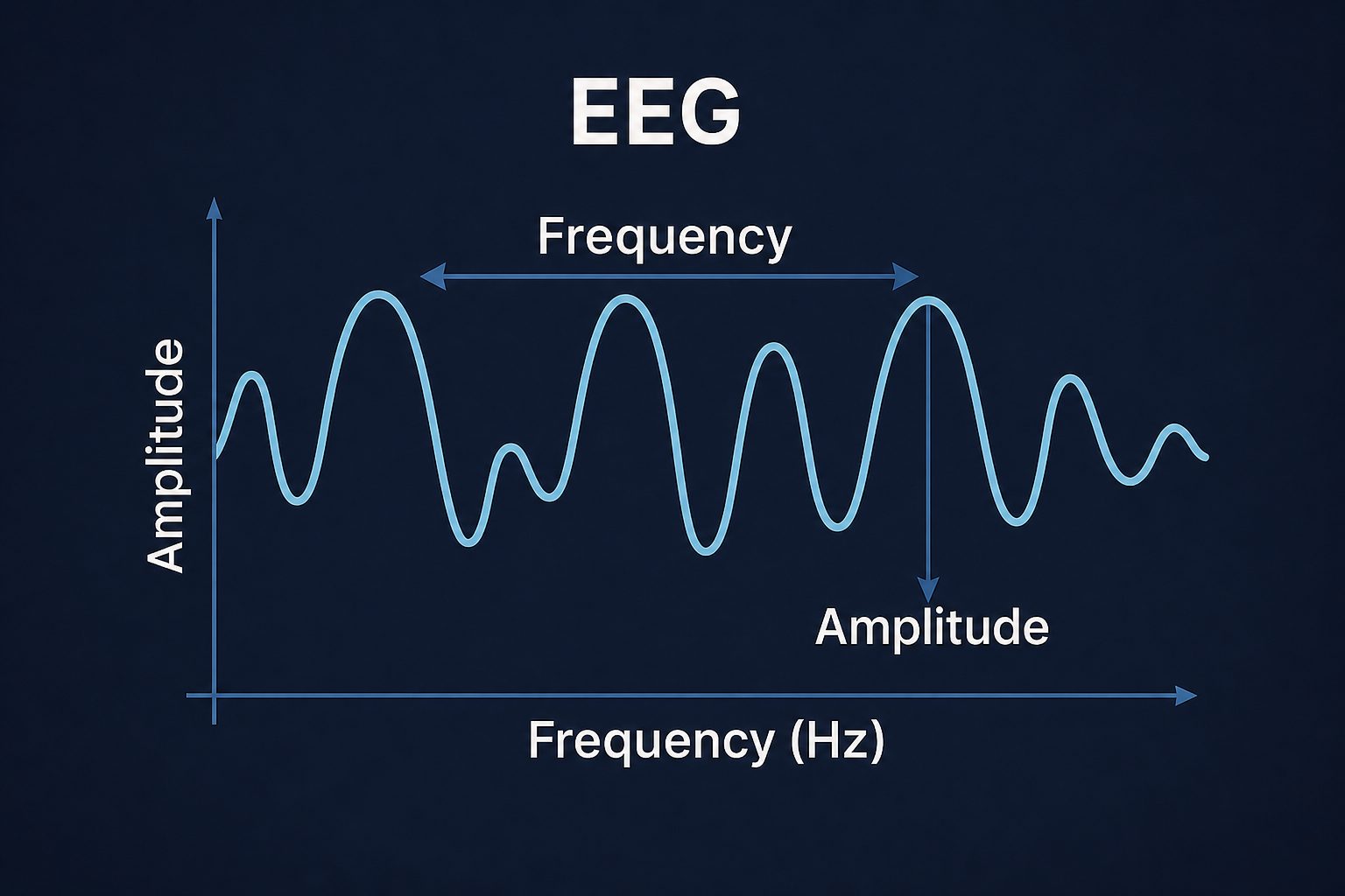
Researchers examine frequency (how quickly a pattern repeats), amplitude (the size of the recorded voltage change), location (where activity is most prominent) and reactivity (how it changes with sleep, eye opening, movement, a task or a stimulus).

Delta-to-Gamma labels summarize parts of a continuous spectrum. Their cutoffs are not universal, and age, alertness, medication, task, recording equipment and analysis choices can affect an EEG. A "brainwave state" means that a pattern is relatively prominent in a recording and context—not that the whole brain has switched to one frequency.

EEG diagram showing frequency and amplitude on a waveform
EEG interpretation depends on frequency, amplitude, timing, location and recording context.

Comparison

The five main brainwave types at a glance

These human brainwave frequency ranges are practical conventions, not rigid boxes. Always check how a study defines its bands before comparing results.

BandApproximate frequencyCommonly observed alongsideInterpretation precautions
Delta0.5–4 HzDeep non-REM sleep; slow activity also occurs in other contextsDelta alone does not prove recovery, healing or sleep quality. Slow signals can also reflect artifacts.
Theta4–8 HzDrowsiness and early sleep; memory and cognitive-control tasksIts presence does not automatically mean dreaming, creativity, hypnosis or subconscious access.
Alpha8–13 HzRelaxed eyes-closed wakefulness, especially over posterior regionsMeaning depends on location, task and reactivity; Alpha is not simply an "idle" signal.
Beta13–30 HzAwake cognitive and sensorimotor processingMore Beta does not automatically mean better focus; muscle tension can add fast activity.
GammaAbove 30 HzFast local activity studied in perception, attention and memoryScalp Gamma is vulnerable to muscle artifact and is not a marker of insight or intelligence by itself.

0.5–4 Hz

Delta brain waves

Delta is the slowest of the five standard bands. In healthy adults, prominent high-amplitude slow activity is a defining feature of deep non-REM sleep, while smaller amounts of Delta-range activity may occur at other times and in specific regions.

This is why Delta is often called the "deep sleep" band. Yet an EEG band is not a benefit by itself. Detecting Delta does not demonstrate physical repair, immune support, emotional integration or restorative sleep. Sleep assessment uses several signals and features, not one frequency alone.

For evening audio, Delta-oriented sleep preparation is more accurate than "activating Delta" or guaranteeing deep sleep. A calm routine may serve as a personal wind-down cue, but responses vary.

What EEG can show

Slow activity can be prominent during deep non-REM sleep under defined recording conditions.

4–8 Hz

Theta brain waves

Theta activity is commonly seen as wakefulness gives way to drowsiness and during early sleep.

Theta-range activity is also studied during waking tasks involving memory and cognitive control, but findings depend on region, task and measurement method. Popular descriptions often equate Theta with dreaming, intuition, creativity, hypnosis or "subconscious access." Theta power alone cannot establish any of those experiences.

Meditation research illustrates the need for context. A systematic review found that Alpha and Theta increases were common in mindfulness studies, but not uniform; methods, experience levels and comparison conditions differed considerably. Continue with Brainwave Frequencies and Meditation.

Theta is also relevant to the transition into sleep, when imagery and dream-like experiences may occur. For the experiential—not diagnostic—side of dream awareness, see Lucid Dreaming: Let's Talk.

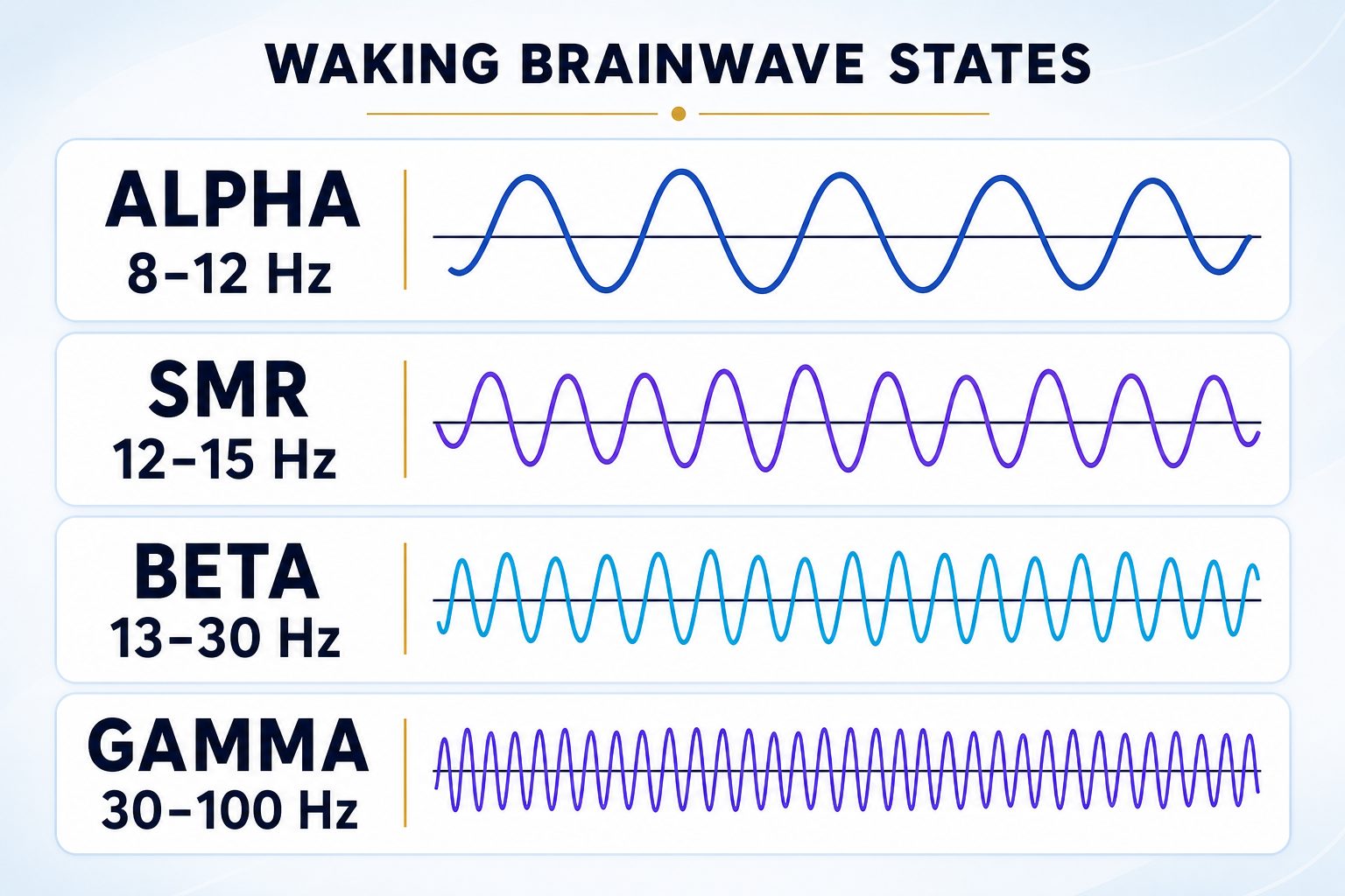
Comparison of Alpha, SMR, Beta and Gamma brainwave frequencies and waveforms
Frequency alone does not determine what a rhythm means; location, task and reactivity also matter.

8–13 Hz

Alpha brain waves

Alpha is one of the clearest rhythms in a normal waking EEG.

In many adults, Alpha becomes prominent over posterior scalp regions when the person is awake, relaxed and has their eyes closed. It usually decreases when the eyes open or attention shifts toward visual input.

This shows why region and reactivity matter. Alpha is not a single whole-brain "calm frequency." Activity near the same frequency over the sensorimotor cortex may be classified as the Mu rhythm rather than posterior Alpha.

Alpha has been studied during attention, sensory processing and meditation as well as rest. It can accompany calm wakefulness without guaranteeing calm, flow, visualization, learning or creativity.

13–30 Hz

Beta brain waves

Beta describes faster activity commonly present during wakefulness.

Beta is studied in active thinking, attention and sensorimotor processing. Its distribution differs from posterior Alpha and can change around movement or motor preparation.

Calling Beta the "focus band" is too simple. Attention relies on networks and changing brain wave patterns across several frequencies. Higher Beta does not prove better concentration, and lower Beta does not prove inattention. Muscle tension and some medications can also affect fast EEG activity.

Beta-oriented audio should therefore be understood as designed for an alert listening context, not as a switch that guarantees concentration or cognitive performance.

Above 30 Hz

Gamma brain waves

Researchers study Gamma-range synchronization and power in relation to perception, attention, memory and communication within or between neural populations. Different studies use different upper limits and may divide Gamma into lower and higher sub-bands.

Gamma findings require particular care. Facial, jaw, neck and scalp muscles generate fast electrical activity that overlaps the Gamma range. Researchers use artifact rejection, task controls and sometimes intracranial recordings to separate neural signals from noise.

Gamma has been reported in some meditation research, but it is not a biological measure of insight, higher consciousness or spiritual development. A frequency band cannot capture the meaning or quality of a person's experience.

Fast and local

Gamma often concerns local, task-dependent activity rather than one uniform whole-brain state.

Artifact risk

Muscle activity can resemble scalp Gamma, so clean recording and careful analysis are essential.

Coexisting rhythms

Can Alpha and Theta brain waves occur at the same time?

Yes. An EEG contains a mixture of frequencies rather than one exclusive wave.

Alpha and Theta can occur during the same recording, with different strengths in different regions or moments. Researchers may compare the power of each band at several electrodes and observe how the patterns change over time.

Brain rhythms can also interact. Cross-frequency coupling research examines whether the phase or amplitude of one rhythm relates to another, and these relationships vary by brain area and task.

Saying that an activity "activates Alpha and Theta at the same time" is an oversimplification. A more precise claim would describe measured changes in Alpha- and Theta-band power or coordination under defined conditions—without implying a universal method or guaranteed experience.

Natural and driven activity

Brainwave entrainment versus naturally occurring brain rhythms

Naturally occurring brain rhythms arise from ongoing neural activity and change with sleep, alertness, sensory input, movement and cognitive demands. Brainwave entrainment concerns the brain's response to periodic external stimulation.

A neural response that follows the timing of an auditory stimulus is not necessarily the same as shifting the brain into a natural sleep, meditation or attention state. Researchers distinguish time-locked auditory responses from broader changes in spontaneous EEG power, connectivity or behavior.

Evidence is mixed. A 2023 systematic review found inconsistent EEG results across binaural-beat studies and substantial differences in protocols, samples and analysis. Rhythmic audio may influence auditory and neural activity under some conditions, but a 10 Hz beat cannot be said to reliably "put the brain into Alpha."

Binaural beats

Each ear receives a slightly different tone. Stereo separation—and therefore headphones—is required for the binaural effect.

Monaural beats

Two tones are combined before playback, so physical amplitude modulation is present in the sound itself.

Isochronic tones

A sound is switched on and off, or strongly modulated, at a regular rate.

Scientific caution

Hearing a beat at a chosen rate is not proof that the brain has entered the corresponding natural state.

Evidence and limits

What scientific evidence can support—and what it cannot

EEG is a powerful measurement tool, but frequency-band labels do not explain a complex mental state by themselves.

Evidence can support

Recurring electrical patterns can be summarized by frequency, amplitude, location and timing.

Context matters

Some patterns are associated with eyes-closed wakefulness, drowsiness, sleep stages or specific tasks.

Bands coexist

Relative power varies across scalp locations and time, and rhythms can interact.

Sound responses vary

Rhythmic sound can evoke measurable responses, while wider entrainment effects vary by protocol.

EEG alone cannot show that one frequency caused an emotion, thought or insight; that a band represents one universal state; or that a sound "activated" the same pattern throughout the brain. It cannot prove creativity, intelligence, spiritual depth or subconscious access, and it cannot show that a listening session heals tissue, rewires the brain or guarantees sleep, focus or meditation.

Scalp EEG has excellent timing resolution but limited spatial detail. Eye movements, muscles, electrode contact and analysis decisions can affect the result. Strong conclusions require suitable controls, reproducible methods and evidence beyond a visually appealing brainwave chart.

Wellness context

Practical, non-medical listening considerations

Use a brainwave label as a design direction, not a promised neurological outcome.

Choose a safe context

Do not use relaxing or sleep-oriented audio while driving, cycling in traffic or doing anything that requires full attention.

Keep volume comfortable

Louder sound does not mean stronger or more effective entrainment.

Check the method

Use stereo headphones when a session specifically relies on binaural beats. Other formats may work through speakers.

Avoid forcing a state

Notice your experience without treating the named band as a target you must achieve.

Stop if uncomfortable

End the session if the sound causes discomfort, agitation, headache or dizziness.

Not medical care

Wellness audio does not replace medical assessment, diagnosis or treatment.

For more on rhythm, texture, intensity and context, read How Sound Affects the Body and Brain. For contemplative practice, explore Meditation and Relaxation.

References

Scientific and institutional sources

The ranges, EEG explanations and interpretation limits in this guide are grounded in the following verifiable sources.

Frequently asked questions

Frequently asked questions about brain waves

What are brainwaves?

Brainwaves are recurring patterns within electrical activity recorded from the brain. Scalp EEG detects voltage changes produced largely by coordinated activity in populations of cortical neurons.

What are the five main types of brain waves?

The five commonly discussed brainwave types are Delta, Theta, Alpha, Beta and Gamma. Their approximate ranges are 0.5–4, 4–8, 8–13, 13–30 and above 30 Hz, although boundaries vary by source.

What is a brainwave state?

It is an informal way to describe a pattern that is relatively prominent in a particular context. It does not mean that only one frequency is present or that the whole brain is uniform.

Do different brain waves control different mental states?

No single band controls one mental state. Associations depend on location, timing, task, alertness and the other activity present in the recording.

What are normal human brainwave frequencies?

Human EEG contains activity over a broad, continuous spectrum. Delta through Gamma are conventional categories, not a checklist of frequencies that must reach one fixed value.

Can several brainwave bands be active at once?

Yes. Multiple frequency components are normally present at the same time, and their relative power varies across scalp locations and over time.

Can Alpha and Theta occur together?

Yes. Alpha- and Theta-band activity can appear in the same EEG recording. Their coexistence does not by itself identify meditation, creativity or a special state of consciousness.

Which brain wave is best for sleep?

There is no single wave to activate. Delta-range slow activity is strongly associated with deep non-REM sleep, while Theta and Alpha changes occur during the transition toward sleep. Delta-rate sound does not guarantee deep sleep.

Which brain wave is best for focus?

Focus cannot be reduced to one frequency. Beta, Alpha, Theta and Gamma can all change during attention tasks, and no audio frequency can guarantee concentration.

Can music or binaural beats change brain waves?

Sound can evoke measurable auditory responses, and some studies report EEG changes during rhythmic stimulation. Binaural-beat findings are inconsistent, and hearing a chosen beat rate does not prove entry into the corresponding natural state.

EEG spectrum Several frequencies coexist.
Regional variation Activity differs across brain areas.
Careful language Association does not prove causation.
No guaranteed state Individual responses vary.

From explanation to experience

Explore a brainwave-oriented listening experience

Choose by context—such as meditation, alert listening or winding down—and approach the session as structured audio rather than a guaranteed neurological result.

Explore Infinity Waves
cierreinterruptor de alimentaciónampliarcruz linkedin facebook pinterest youtube rss twitter instagram facebook-blank rss-blank linkedin-blank pinterest youtube twitter instagram